2-[18F]FDG PET is the standard in clinical molecular imaging but relies on radiation, detects only one metabolic phenotype, and often misses cancers with atypical metabolism, such as RCC. Fluorine Metabolic Imaging (FMI) uses nonradioactive fluorinated saccharides like 3-[19F]FDGal as probes to map the enzymes AKR1B1 and ALDH1A1 via MRI. The method enables simultaneous visualization of the probe and its products, such as 3FDGtol, which reports on AKR1B1 activity and highlights RCC lesions with background-free 19F-MRI. By replacing 19F with the 18F isotope, the same approach can be adapted for PET, combining FMI’s metabolic specificity with the high sensitivity of standard PET systems.
- MRI-based molecular imaging of various cancers, including RCC
- Companion diagnostic for ALDH1A1- or AKR1B1-targeted therapies (for oncological and non-oncological indications, i.e., Epalrestat treatment diabetic neuropathy)
- Broad cancer metabolic imaging, potentially patient-tailored
- Functional characterization of tumor metabolism
- Preclinical research tool for drug development for RCC, metabolic diseases, and others
- Enzyme-based molecular imaging. Also fits RCC biology
- Dual-modality: Compatible with 19F-MRI and 18F-PET
- High Resolution with no background signal from soft tissues
- Non-invasive and organ-specific
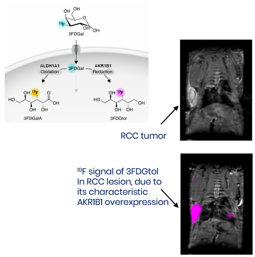
Demonstrated in vivo proof-of-concept using murine xenograft models of NSCLC and RCC. Robust 3FDGal uptake and metabolic conversions to 3FGtol (via AKR1B1) and to 3FDGalA (via ALDH1A1), confirmed in vitro and in vivo with NMR, MRI, and multimodal validations. Additional fluorinated sugar analogs also show promise. PET adaptation remains to be validated.


Dr. Vered Pardo Yissar