Standard light microscopy generates flat, two-dimensional images of transparent specimens, making it difficult to resolve the spatial arrangement of internal structures. Existing methods to infer depth rely on multiple recordings or complex hardware adjustments, often resulting in inaccuracies. This technology introduces a novel single-scan method for obtaining both phase and depth contrast in transmission microscopy by using a multi-segment detector and a proprietary parallax correction algorithm, enabling more precise and efficient analysis without complex setup or multiple acquisitions.
- Light microscopy of transparent samples
- Confocal laser scanning microscopy
- Cell biology, neuroscience, and developmental biology
- Materials science and nanotechnology
- Semiconductor and microelectronics imaging
- Depth and phase contrast from a single scan
- Eliminates the need for multiple focus-level recordings
- Reduces artifacts caused by image alignment errors
- Simplified optical setup
- Enhanced accuracy in interpretation of 3D structural information
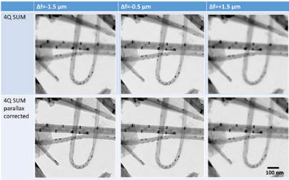
Focus extension in the sum images of OPAL bright-field quadrants by compensating the parallax image shifts
Demonstrated in both electron and light microscopy. Functionality was validated using a quadrant detector and a parallax correction algorithm, which together enhance image contrast and depth interpretation.