16721
Overview
An advanced deep-learning method for analyzing super-resolution Ultrasound Localization Microscopy (ULM) data to quickly and accurately reconstruct microvasculature structures, without needing prior knowledge of the system's characteristics. This enables improved US imaging for early diagnosis and clinical management of various diseases, including breast cancer and Crohn's disease.
Applications
- Microvascular imaging using super-resolution US for clinical use
- Distinction between different types of breast tumors
- Early diagnosis and monitoring of various diseases characterized by microvasculature alterations
Differentiation
- Fast Analysis
- Radiation free
- Cost effective
- Accessible
- No prior knowledge of system parameters (PSF or UCAs) required
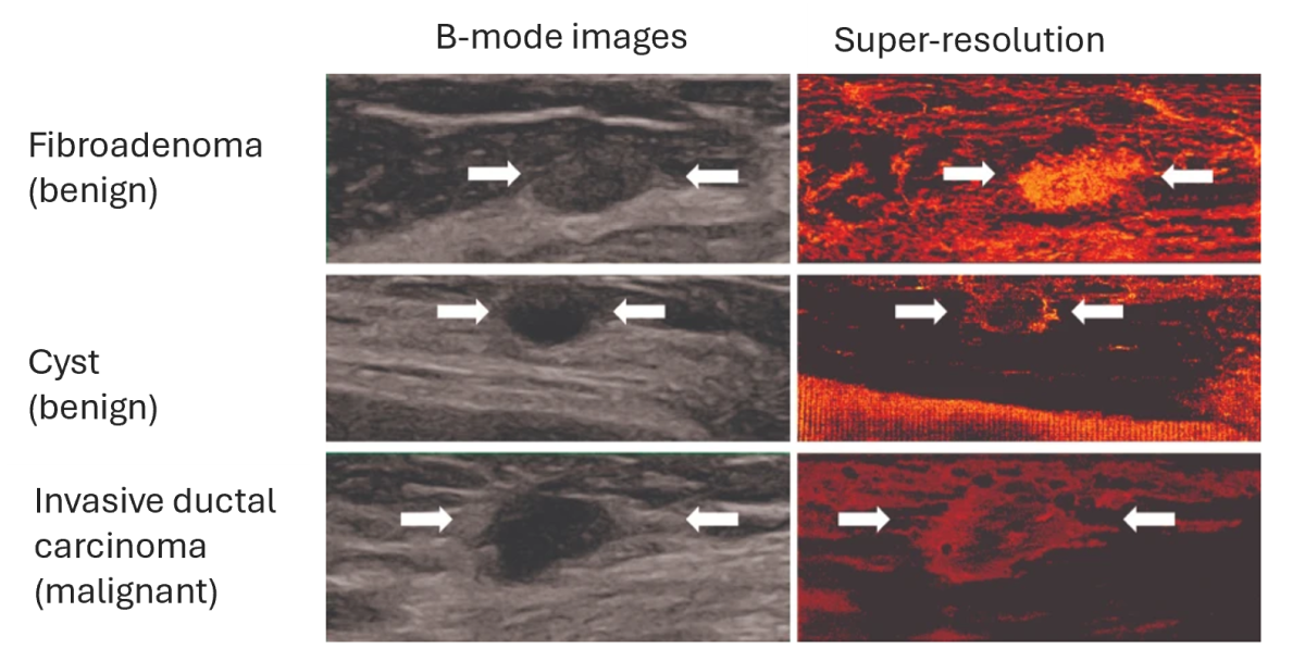
Comparison of conventional B-mode and super-resolution US images of three breast lesions
Development Stage
The method has been clinically validated in breast cancer patients and is being further tested for other applications. A US patent application has been published.
Patent Status:
USA Published: Publication Number: US 2024/0249384

Contact for more information